Treatment for Brain Aneurysms

What is an aneurysm?
An aneurysm occurs when a section of an artery wall weakens, causing the artery to abnormally widen or bubble out as blood circulates through the weakened artery.
An aneurysm in the brain is called a cerebral aneurysm.
What causes an aneurysm?
There is no one specific cause of an aneurysm. Some are genetic, while others may form as a result of injury or other unknown factors.
While a definitive cause of most aneurysms is unknown, there are several risk factors that can contribute to weakening of arterial walls and therefore, the formation of an aneurysm. Significant risk factors include high blood pressure, smoking, and drug and alcohol abuse.
How is an aneurysm diagnosed?
Play Podcast
Download Podcast
Brain aneurysms can be present without any symptoms and, as a result, may go undetected for a long period of time.
Symptoms and intensity also vary depending on whether the aneurysm has ruptured, is stable or leaking:

Anyone experiencing any combination of these symptoms should go to the emergency room immediately.
A series of tests can help determine whether an aneurysm is present and whether it has ruptured.
Tests may include:
Cerebral angiogram. With the help of biplane interventional imaging, a small tube called a catheter is inserted into a major artery (typically, in the groin) and guided through the arteries in the heart up to the brain.
Biplane imaging technology produces detailed 3-D images of of the structure and location of blood vessels, soft tissues and blood flow in real-time, enabling providers to determine if an aneurysm is present and evaluate its condition.
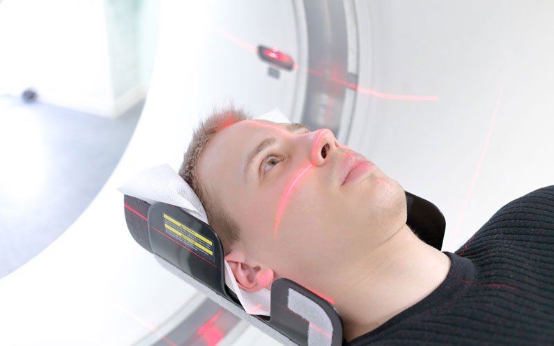
CT scan (computerized tomography). This scan is performed to identify bleeding in the brain, and is often the initial step in diagnosis. The scan produces images similar to an X-ray, but CT technology captures enhanced 2-D images of the brain, as opposed to a one-dimensional, single X-ray image.
Contrast dye is often injected into the blood vessels to help highlight blood flow throughout different areas of the brain, better enabling providers to detect any abnormalities, such as an aneurysm. When dye is used to assist in this type of scan, the variation is called a CT angiogram.
MRI (magnetic resonance imaging). An MRI renders intricate images of the brain, utilizing powerful magnets and radio waves, rather than radiation, to produce 2-D and 3-D images of the soft tissue. Differences between normal and abnormal tissue are often more clear on an MRI image than on a CT image, which uses X-rays.
Spinal tap (Cerebrospinal fluid test). If an aneurysm has ruptured, red blood cells likely will be found in the fluid surrounding the spine and the brain (cerebrospinal fluid).
This test, also called a lumbar puncture, is often performed if a patient presents symptoms of an aneurysm but the CT scan images do not provide enough evidence for a definitive diagnosis. Using a needle, the provider draws fluid from the patient’s back and tests it for red blood cells.
What are the treatment options for an aneurysm?
EvergreenHealth uses two primary methods of treatment to repair brain aneurysms: coiling and clipping.
Clipping, or open repair, involves placing small metallic clips along the blood vessel that feeds the aneurysm, which stops the blood flow to it. Because part of the skull must be removed to perform this surgery, the recovery time is typically four to six weeks.
Endovascular coiling, also called embolization, is a minimally invasive procedure made possible with the use of our biplane interventional imaging.
The detailed, 3D images produced by the biplane help the provider guide a catheter containing a metal wire into the aneurysm through the vascular system (typically through an artery in the groin) where it coils up and stops the blood flow, sealing off the aneurysm from the artery.
Because coiling is a less invasive procedure, those patients face a shorter recovery time—only two to four weeks—and can leave with little evidence the procedure has been done.